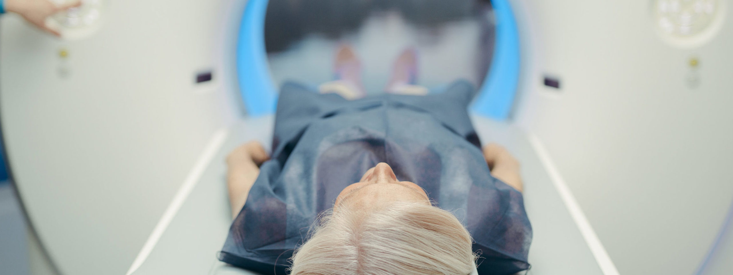
Diagnostic imaging tests: The Difference between XRay, UltraSound, MRI, CT scan
There are numerous specific types of diagnostic imaging tests. Each creates images primarily based totally on different technology. A health practitioner can also additionally order one, or more than one test so that it will assist diagnose or rule out certain medical complications. Some of the maximum common types of diagnostic imaging tests besides X-rays are magnetic resonance imaging tests (MRIs), Computed Tomography scans (CT), and Ultrasound.
Unfortunately, the thought of having those tests done can regularly make patients anxious, however, it’s important to remember that diagnostic imaging is commonly non-invasive and painless. Therefore, in case your health practitioner recommends which you are in want of specialized diagnostic imaging, it is able to be beneficial to recognize each how they work, and the common makes use of for the specific styles of imaging. Knowing the variations of every of those imaging tests can assist ease your thoughts and know what to expect. Here are a number of the most common tests we provide at Independent Imaging:
- X-rays (radiographs) are the maximum common and broadly to be had diagnostic imaging technique. Bones, calcifications, a few tumors, and different dense matter seem white or mild due to the fact they absorb the radiation. Less dense tender tissues and breaks in bone permit radiation pass through, making those elements appearance darker on the x-ray film.
- Ultrasound generation is a great diagnostic device for seeing live images of the working structures of the body, especially the structures of joints within the body. Ultrasound imaging (sonography) makes use of excessive frequency sound waves, to create a live video feed picture of the inner of the body. Ultrasound is the generation, or the “eyes” if you will, for supporting docs get a more in-depth appearance to make an accurate diagnosis. Since images are captured in real-time all through ultrasound, they also can display the movement of the body’s internal organs in addition to blood flowing through blood vessels. Unlike X-ray imaging, there may be no radiation exposure associated with ultrasound imaging.
- An MRI makes use of an effective magnetic field mixed with specific radio frequencies to create specified pix of inner frame systems with the useful resource of an advanced computing system. MRI’s can stumble on abnormalities, cancerous and noncancerous growths, broken tissues, and greater. They also can assist your health practitioner advantage a higher know-how of your joints, cartilage, bone, and tender tissues in a manner that different assessments cannot.
- A computed tomography scan referred to as a CT experiment combines a chain of X-ray images taken from many specific angles with computer processing generation, to create sections of images of the bones and tender tissue in the body. For example, a CT scan may be associated with reducing bread. Each segment or slice of images may be regarded in my view to get a higher visualization and universal image of the body. CT scan images can offer a good deal greater facts than simple X-rays and other diagnostic imaging.
Photo by National Cancer Institute on Unsplash


