Cone beam computed tomography in Endodontics
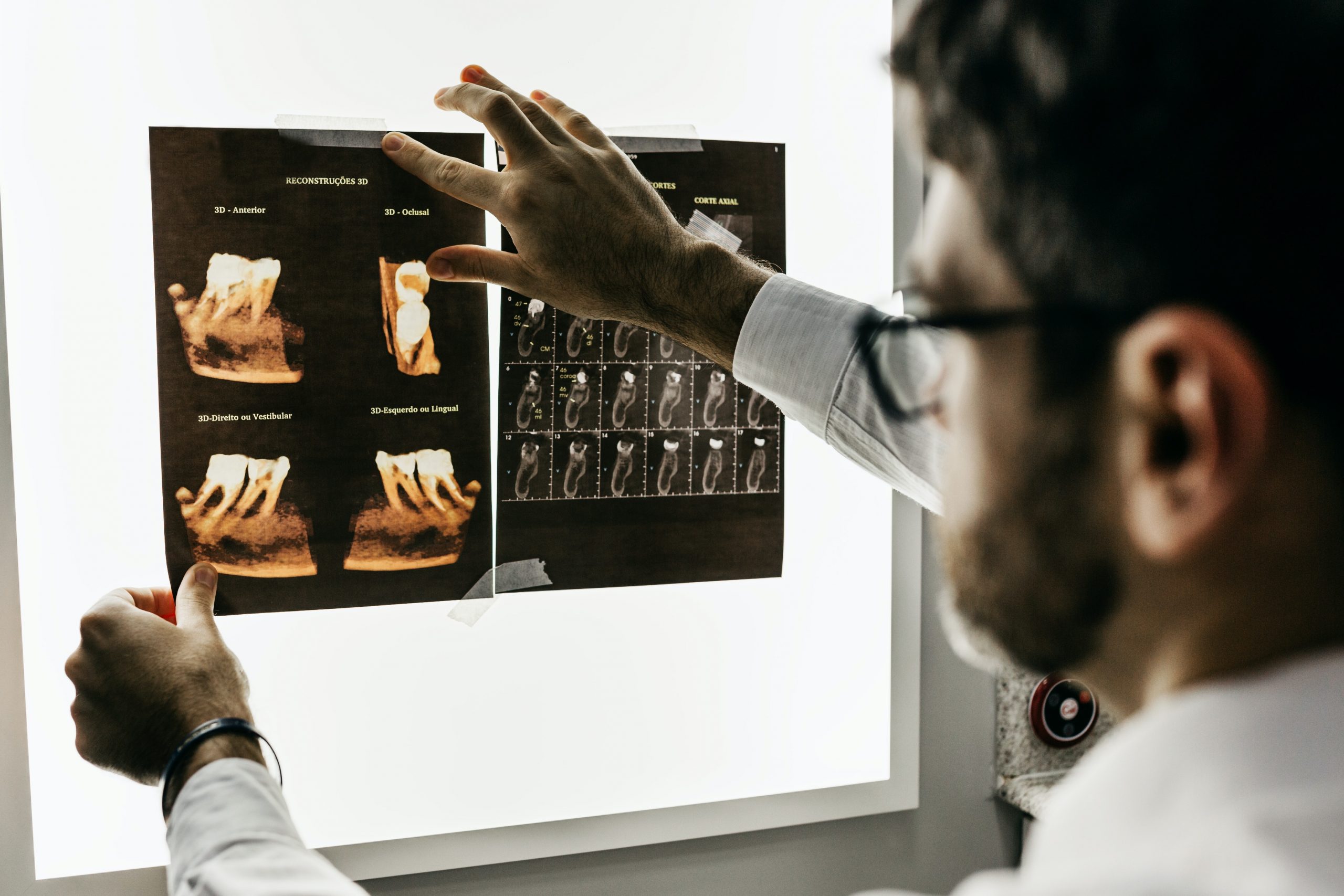
Cone Beam Computed Tomography in Endodontics is a three-dimensional imaging technology that has revolutionized the field of endodontics. It offers higher resolution and lower radiation exposure compared to conventional computed tomography (CT) and provides accurate imaging of the root canal system.
Diagnosis and Treatment Planning
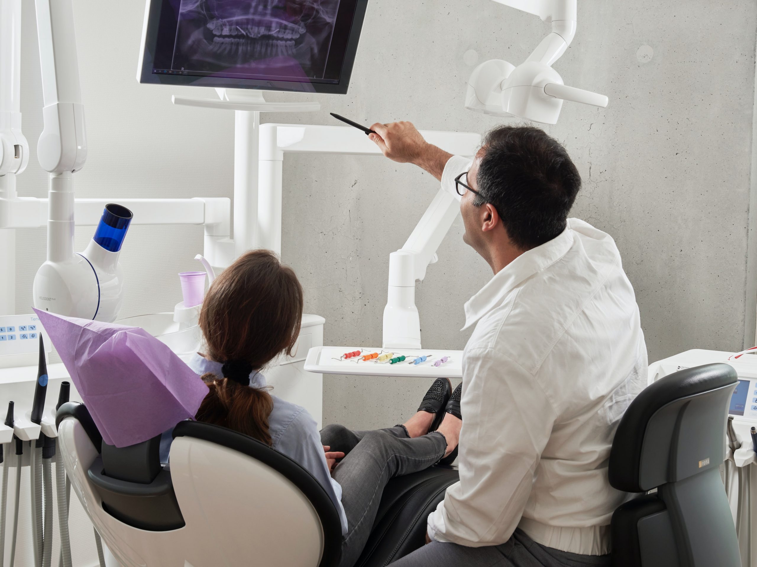
Cone beam computed tomography(CBCT) is a valuable diagnostic tool for endodontists, enabling them to accurately diagnose and plan treatment for complex endodontic cases. CBCT images provide a three-dimensional view of the teeth and their surrounding structures, allowing for a more accurate diagnosis of root canal anatomy, periapical pathology, and tooth fractures. This helps endodontists to determine the best course of treatment, including the need for surgical intervention, root canal re-treatment, or tooth extraction.
Identifying Root Canal Anatomy
The complex anatomy of the root canal system is a significant challenge in endodontics, making it challenging to detect and treat all canals effectively. CBCT imaging offers high-resolution, three-dimensional images that allow for accurate detection of extra canals, lateral canals, and other anatomical variations. This information is essential for accurate treatment planning and successful outcomes in endodontic therapy.
Surgical Endodontics
Computed tomography in endodontics, such as apical surgery and guided tissue regeneration. CBCT images provide a more accurate depiction of the root apex, periapical region, and surrounding bone, allowing endodontists to identify and treat pathology more precisely. This can help to minimize the surgical trauma and reduce the risk of complications.
Implant Planning
CBCT is also useful in implant planning and placement. CBCT images provide accurate measurements of bone density, quality, and volume, allowing for precise implant placement and optimal implant angulation. This can result in a better functional and aesthetic outcome, reducing the risk of implant failure and bone resorption.
Radiation Exposure
CBCT does expose patients to ionizing radiation, although the dose is much lower than conventional CT imaging. The risk of radiation exposure should be carefully weighed against the benefits of CBCT imaging, and the ALARA principle (As Low As Reasonably Achievable) should be followed to minimize radiation exposure.
Conclusion
CBCT imaging has revolutionized the field of endodontics, providing accurate and detailed three-dimensional imaging of the root canal system. It has numerous applications in diagnosis, treatment planning, identifying root canal anatomy, surgical endodontics, and implant planning. However, the risk of radiation exposure should be carefully considered and minimized whenever possible. With proper usage and interpretation, CBCT can significantly improve the outcomes of endodontic treatment.


