
X-ray Dark-Field Imaging can Detect and Quantify Emphy-sema in COPD Patients
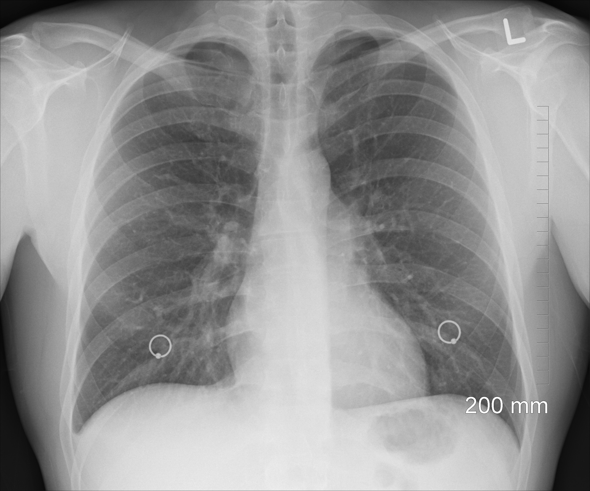
Diseases of the respiration system are the main international reasons for persistent morbidity and mortality. While superior scientific imaging technology these days supply special diagnostic data, a low-dose, fast, and less expensive choice for early detection and/or follow-ups continues to be lacking. It was reported the first human application of a unique modality, particularly X-ray dark-field chest imaging, may fill this gap. Enabling the evaluation of microstructural adjustments in the lung parenchyma, this approach provides a more sensitive alternative to traditional chest X-rays, and but calls for handiest a fragment of the dose applied in computed tomography (CT).
Until recently, the use of dark-field X-rays imaging had been studied in small animals and cadavers, however never humans. That is till researchers at the Technical University of Munich (TUM) put the generation to test with patients. They determined that dark-discipline imaging may be a treasured and cost-powerful device in the early analysis of respiration diseases. For the study, researchers at TUM designed and built a novel dark-field chest X-ray system able to produce typical thorax radiographs, focusing on the early detection of emphysema in sufferers with COPD. A general of seventy-seven sufferers who had gone through medically indicated chest CTs has been recruited. Those with situations apart from COPD that would affect lung parenchyma have been excluded. Five separate readers received and reviewed chest CTs and dark-field X-rays for all sufferers. Compared to the CT scans, the dark-field sign yielded a more potent correlation with lung diffusion capacity. In addition, the dark-field emphysema evaluation became steady with the outcomes of the CT scans, however, without the extended radiation dose, CT imaging requires. The radiation dose of dark-field imaging may be decreased through an aspect of fifty, according to the study.
X-ray dark-field chest imaging allows the diagnosis of pulmonary emphysema because it affords applicable data representing the structural circumstance of lung parenchyma. Significant diagnostic advantages also are predicted for different lung disorders.